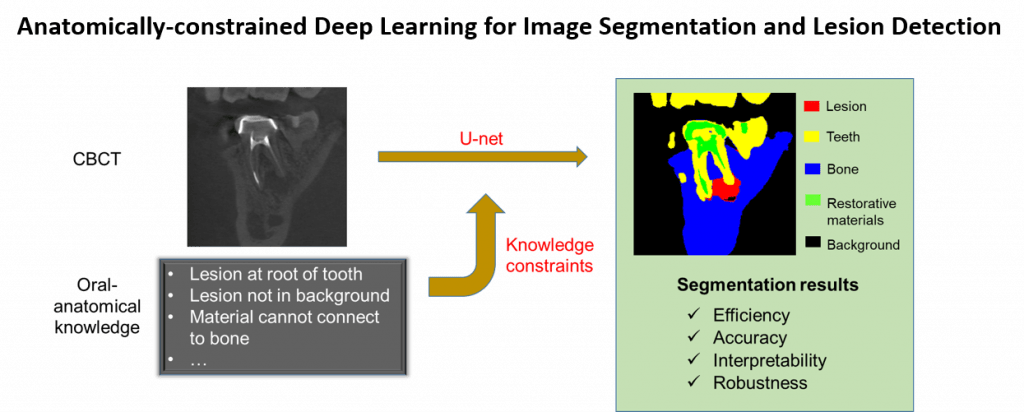
Cone beam computed tomography (CBCT) is a 3D imaging modality that is becoming widely adopted in a variety of dental practices to aid diagnosis and treatment planning of oral diseases. Because the oral environment is structurally very complex and has substantial individual differences, clinician-based CBCT interpretation lacks precision, consistency, and objectivity. Existing computer-aided detection/diagnosis (CAD) algorithms also fall short of resolving the 3D image complexity, thus providing limited clinical utility. Deep learning (DL) algorithms are suitable for image analysis, but pure data-driven learning may be limited by sample size constraints and lack of interpretability. This project aims to innovate DL by integrating domain knowledge of human oral anatomy into the DL design, namely anatomically-constrained DL, to allow for segmentation of CBCT images and detection of lesions with better accuracy, efficiency, and interpretability.
