4D Flow Magnetic Resonance Imaging for the Study of Complex Blood Flow Patterns in Carotid Webs
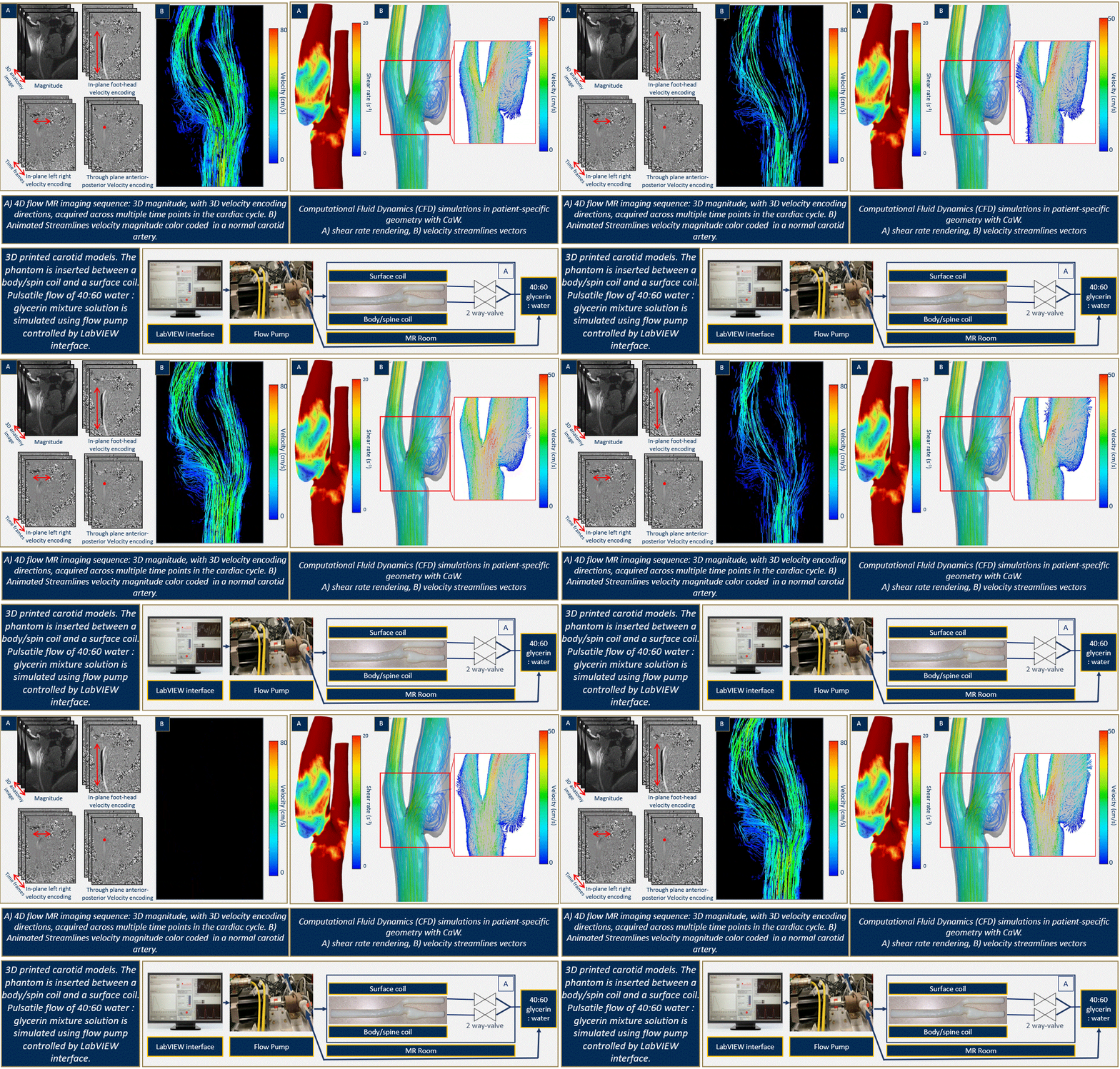
Carotid Web (CaW) accounts for up to 37% of cryptogenic recurrent ischemic strokes in young patients (<55 years old). CaWs have an estimated population prevalence of up to 1-7% (estimated 3.3-23.1 million people in the United States with CaW). Although the risk of stroke in this population is currently unknown, it is critical to identify those at high risk for stroke so that appropriate treatment can be instituted prior to an ischemic event.
Hemodynamic parameters of normal and atherosclerotic carotid artery bifurcations have been investigated previously using experimental and computational modeling studies. Even though CaW generates similar hemodynamics disturbance to that in atherosclerotic lesions, the sudden contraction and expansion created by a CaW can cause an extreme flow disturbance. This can be represented by Larger recirculation zones and lower shear rates are expected in CaW compared to atherosclerotic lesions.
5D Flow MRI & Image Reconstruction

5D free-running image acquisition is a novel non-ECG gated and free-breathing 3D isotropic resolution whole-heart scan. It was leveraged to quantify flow at the same time to 5D flow imaging.
Studying the 3D Changes in Myocardial Deformation via 3D DENSE MRI
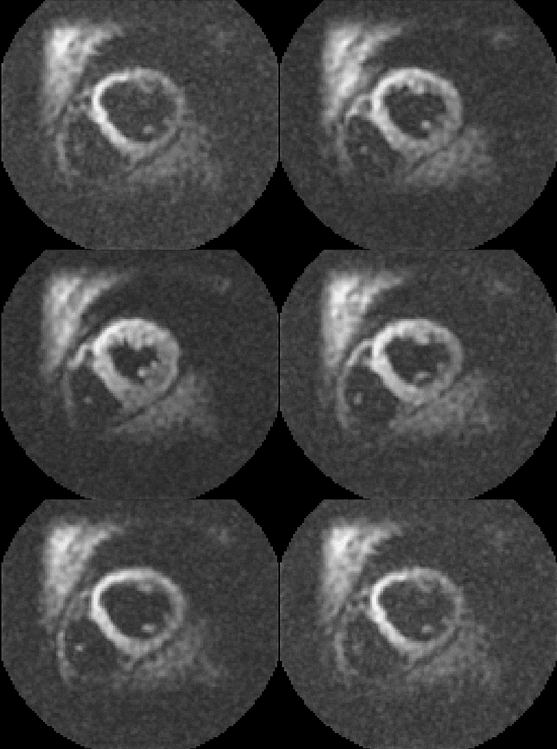
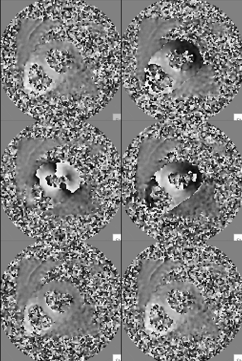
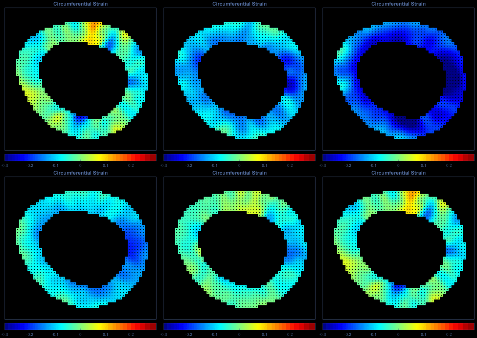
Myocardial deformation (i.e., the changes in its size and shape), lies at the heart of understanding the physiological activity of the heart. Towards that end, we are interested in studying the local changes in myocardial deformation in heart disease, potentially gaining more insights into cardiac pathologies as well as investigating their utility in predicting disease. Thus, displacement encoding with stimulated echoes (DENSE) is an MRI pulse sequence that can estimate the pixel displacement field of the myocardium, which can then be used to calculate the Green strain, a measure of finite deformation, over the course of a cardiac pathology.


Magnetic Resonance Elastography (MRE)
Brain stiffness quantification by applying fixed frequency to the skull during MR acquisition..
